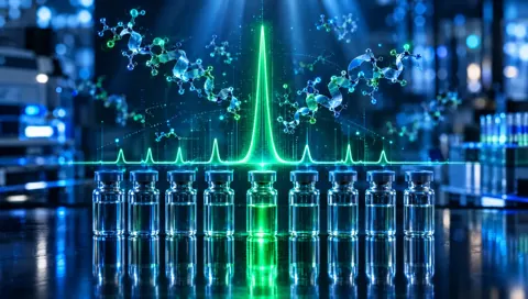
214 nm
Far UV
205–214 nm
Strongest peptide-bond signal and highest sensitivity. More background from solvents and additives, so baselines can drift.
(“U-V WAVE-length”)
One number on the chromatogram that decides what the detector can see.
The short answer
The detection wavelength is the color of ultraviolet light an HPLC detector uses to see each component as it leaves the column; for peptides it is usually 214 to 220 nm, where the peptide bond absorbs.
A UV detector shines light through the liquid leaving the column and measures how much is absorbed. Every peptide bond absorbs strongly around 200 to 220 nm, so detecting there sees every peptide in proportion to its length. At 280 nm only tryptophan and tyrosine absorb much, so peptides without them nearly vanish. The wavelength therefore shapes which impurities are visible and how purity is calculated.
Reviewed by the Codex Research quality teamUpdated Nov 8, 2026
Word study
Say it
ul·tra·vi·o·let wave·length
(“U-V WAVE-length”)
Roots and origin
RootLatin
OriginOld English
Ultraviolet means beyond violet, the shortest wavelength the eye can see. Peptide detection uses light well past that edge, in the far ultraviolet, where the bonds that hold every peptide together absorb strongly.
Around the world
Tap a language to hear it spokenDefinition
An HPLC UV detector works like a very precise light meter. As each separated component flows through a small cell, the detector shines ultraviolet light through it at a set wavelength and records how much is absorbed. The more a component absorbs at that wavelength, the bigger its peak. The chosen wavelength is part of the method and is usually printed on the chromatogram.
Peptides are almost always detected at around 214 to 220 nm. Each peptide bond absorbs in this region, so the signal roughly follows the number of bonds and every peptide in the sample is seen, whatever its sequence. Published measurements put the peptide bond at about 923 M⁻¹cm⁻¹ at 214 nm, with aromatic residues such as tryptophan absorbing many times more.
At 280 nm the picture changes. Only tryptophan and tyrosine absorb strongly, at about 5,500 and 1,490 M⁻¹cm⁻¹ respectively, which is why 280 nm is the standard for measuring protein concentration. A peptide with neither residue is nearly invisible at 280 nm, and a purity result measured there could miss impurities that lack aromatic residues entirely.
The trade-off at low wavelengths is background. Many solvents and additives also absorb below about 220 nm, including the TFA used in many mobile phases, which can make the baseline drift as the gradient runs. Methods balance sensitivity against a clean baseline, which is why 214, 215 and 220 nm are all common.
For a buyer, the wavelength matters because HPLC purity is an area percentage at one wavelength. Components that absorb more strongly look bigger than they are, and components that barely absorb look smaller. A certificate that states the wavelength lets you judge whether the method could see the impurities it claims to have measured.
Common choices
Strongest peptide-bond signal and highest sensitivity. More background from solvents and additives, so baselines can drift.
A common compromise for peptides: strong backbone signal with less interference from mobile-phase additives.
A traditional default on older fixed-wavelength detectors. Weak for most peptides unless they contain aromatic residues.
Dominated by tryptophan and tyrosine. Standard for protein concentration, but blind to peptides that lack them.
A detector that records the whole spectrum at once. It can show whether a peak is spectrally uniform and help spot hidden co-elution.
Interactive
Move the detection wavelength across the ultraviolet, then add a tryptophan or a tyrosine. Without them, the peptide bonds do all the absorbing at 214 nm and almost nothing is left by 280 nm. Aromatic residues absorb strongly in both regions, and at 280 nm they are all the detector sees.
A 15-residue peptide containing
An approximate model built from published molar absorptivities: about 923 M⁻¹cm⁻¹ per peptide bond at 214 nm, and 5,500 for tryptophan and 1,490 for tyrosine at 280 nm. Band shapes are simplified, and the vertical axis is logarithmic so both regions fit on one chart.
For buyers
An impurity that does not absorb at the chosen wavelength does not appear as a peak, however much of it is present.
Purity is an area percentage at one wavelength. Change the wavelength and the relative peak sizes change with it.
Detection near 214–220 nm for peptides is a basic sign that the method was designed for peptides at all.
At Codex Research
Each Codex Research product page carries a lot-matched certificate of analysis reporting HPLC purity and mass spectrometry identity for the lot in stock.
See it live: the HPLC guide in the glossary
Myth vs fact
Tap a card to flip it.
Watch for
Quick check
Printed on the chromatogram or in the method notes.
The usual range for peptide purity.
At 280 nm, the peptide should contain Trp or Tyr.
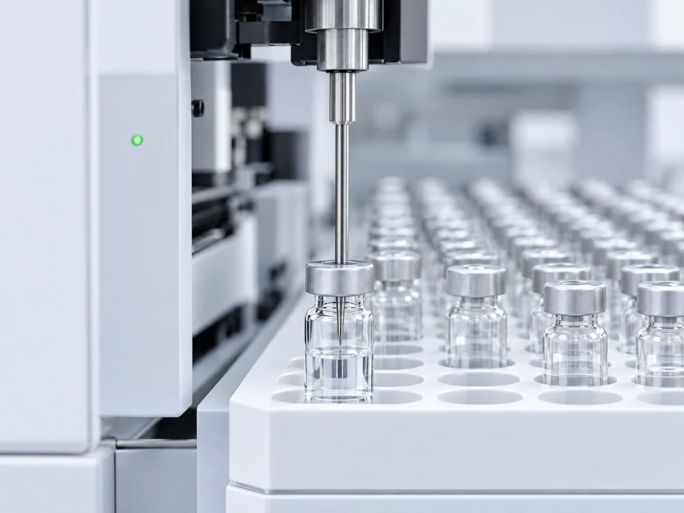 Full walkthrough · JournalHPLC vs mass spec: how purity is tested
Questions
Because the peptide bond absorbs strongly there, so every peptide is seen in proportion to its number of bonds, whatever its sequence.
At 214 nm the backbone absorbs; at 280 nm mainly tryptophan and tyrosine do. Peptides without those residues barely show at 280 nm.
Usually 214 to 220 nm. The wavelength should be stated with the purity result.
Many solvents and additives also absorb below about 220 nm, and their concentration changes during a gradient run.
Yes. Purity is an area percentage at one wavelength, and components absorb differently at different wavelengths.
A UV detector that records the full spectrum for every point in the run, which helps check whether a peak is a single component.
All peptide bonds absorb in the far UV. Near 280 nm, only tryptophan, tyrosine and, weakly, cystine contribute much.
Sources and standards
Written and reviewed by the Codex Research quality team · Last reviewed November 8, 2026 · For laboratory research use only.
On real products
These products’ current certificates report purity by RP-HPLC, the method behind wavelength. See the result on each product page, then read the full guide.
 Cellular
Research peptide
Cellular Research
SS-31 (elamipretide) is a synthetic tetrapeptide that binds cardiolipin at the inner mitochondrial membrane, studied in bioenergetics and oxidative-stress research.
 Blends
Research peptide
Neuropeptide Research
A blend of Semax, an ACTH(4-7) analog, with Selank, a tuftsin analog. Both are seven-residue peptides from the same Russian design programme.
 Neuro
Research peptide
Neuropeptide Research
DSIP is a nine-residue peptide isolated from rabbits in the 1970s and named for the delta EEG pattern it produced, studied in neuropeptide research.
Journal guideHPLC vs mass spec: how purity is testedRead the guideKeep going
 From lab to your door
U.S.-made research vials, shipped with lot-matched, lab-tested COAs you can scan straight off the label.

21+ Only
Codex Research provides research-grade compounds for laboratory and research use only. By entering, you confirm you are at least 21 and agree to our Terms & Conditions.
By proceeding you affirm the statements above are true. Products are supplied for laboratory research use only — not for human or veterinary use, not for use in diagnostic procedures, and not evaluated by the U.S. Food and Drug Administration. Full disclaimer.